
The cell is the fundamental unit of life, and animal cells play a crucial role in the functioning of living organisms. In this article, we will explore the structure, organelles, and characteristics of animal cells. We will also compare animal cells to plant cells to understand their differences. So, let’s dive into the fascinating world of animal cells.
An Introduction to Animal Cells
Animal cells are a type of eukaryotic cells, which means they have a true, membrane-bound nucleus and other membrane-bound organelles. These cells are the building blocks of animals and are responsible for carrying out various functions necessary for life. Animal cells come in a variety of shapes and sizes, ranging from microscopic to millimeters in length. They lack a cell wall, which allows for greater flexibility and adaptability compared to plant cells.
What is an Animal Cell?
An animal cell is a type of eukaryotic cell that lacks a cell wall and has a true, membrane-bound nucleus along with other cellular organelles. These organelles include the cell membrane, nucleus, cytoplasm, cytoskeleton, lysosomes, nucleolus, mitochondria, endoplasmic reticulum, ribosomes, Golgi apparatus, peroxisomes, centrosome, vacuole, nucleopore, cilia and flagella, endosomes, intermediate filaments, microfilaments, microtubules, plasma membrane, and microvilli.
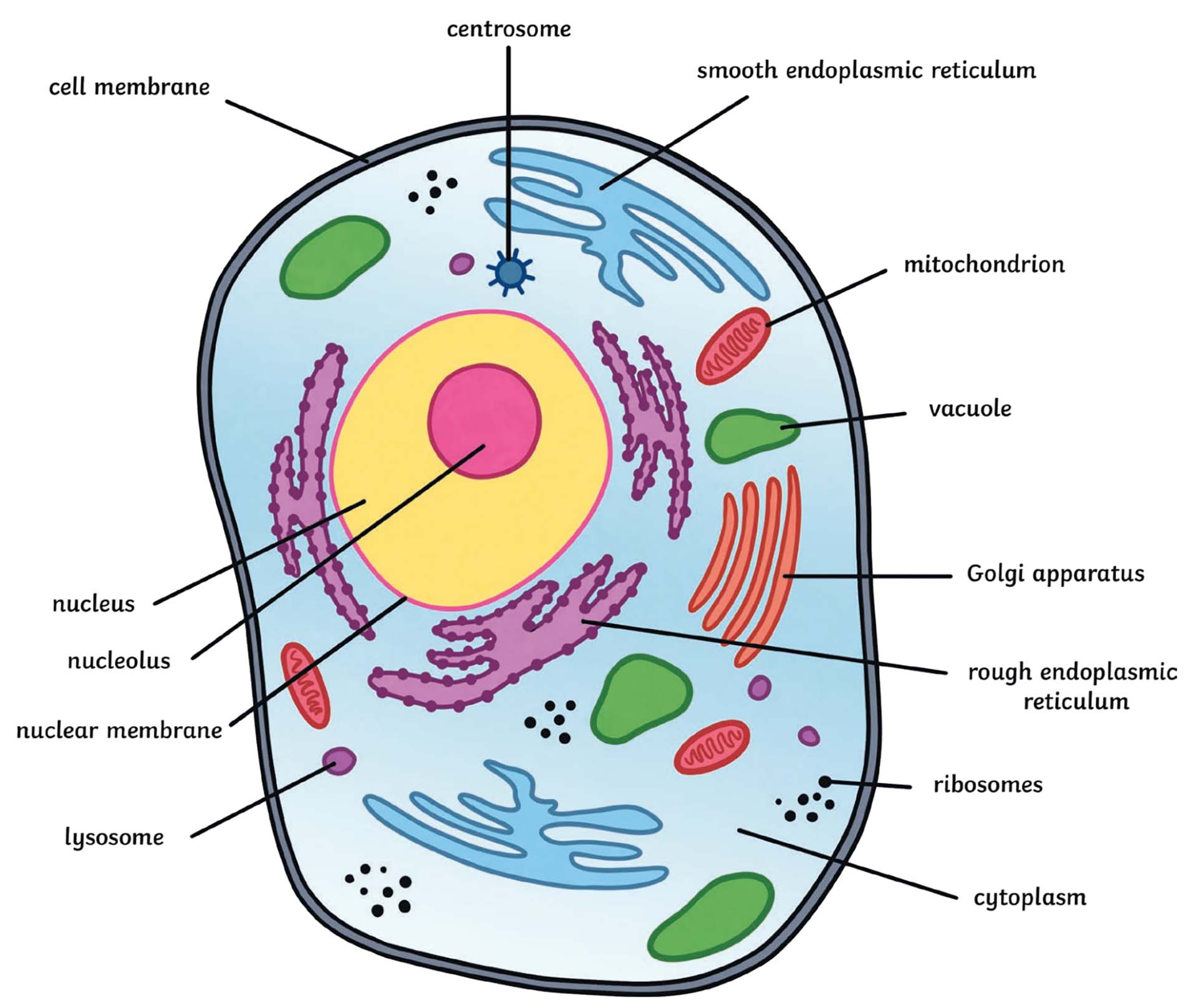
Animal Cell Structure: Organelles and Their Functions
Cell Membrane
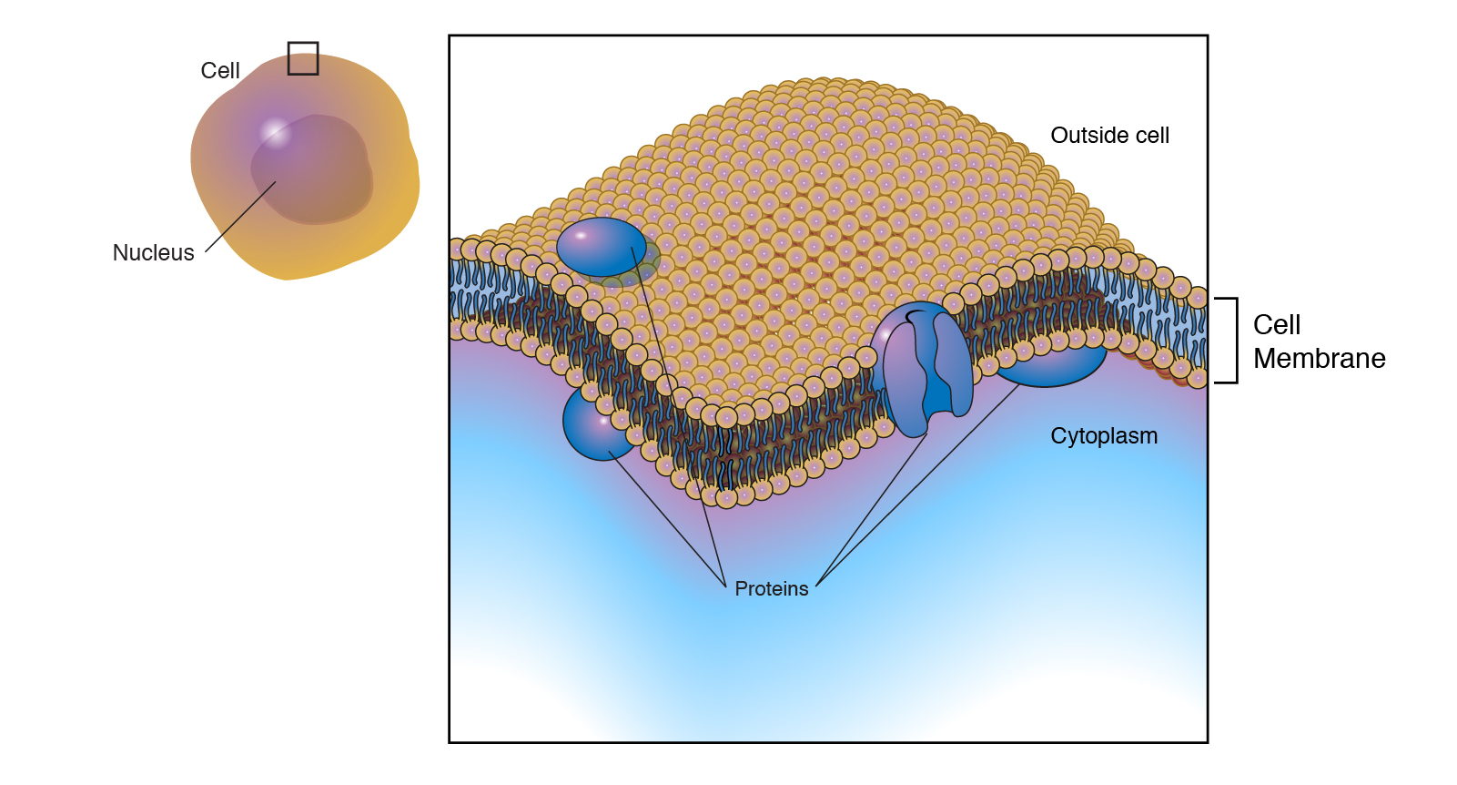
The cell membrane, also known as the plasma membrane, is a thin, semipermeable layer of lipids and proteins that surrounds the animal cell. It acts as a protective barrier, controlling the entry and exit of nutrients and other microscopic entities into the cell. The cell membrane is selectively permeable, allowing only certain substances to pass through.
Structure of Cell Membrane: The cell membrane is composed of a lipid bilayer with embedded proteins. The lipid bilayer consists of two layers of phospholipids, with the hydrophilic heads facing outward and the hydrophobic tails facing inward. The proteins embedded in the lipid bilayer have various functions, including transport of molecules, cell signaling, and cell adhesion.
Functions of Cell Membrane:
- Protects the cell from its surroundings
- Controls the entry and exit of substances into and out of the cell
- Facilitates cell communication and signaling
- Maintains cell shape and structure
Nucleus
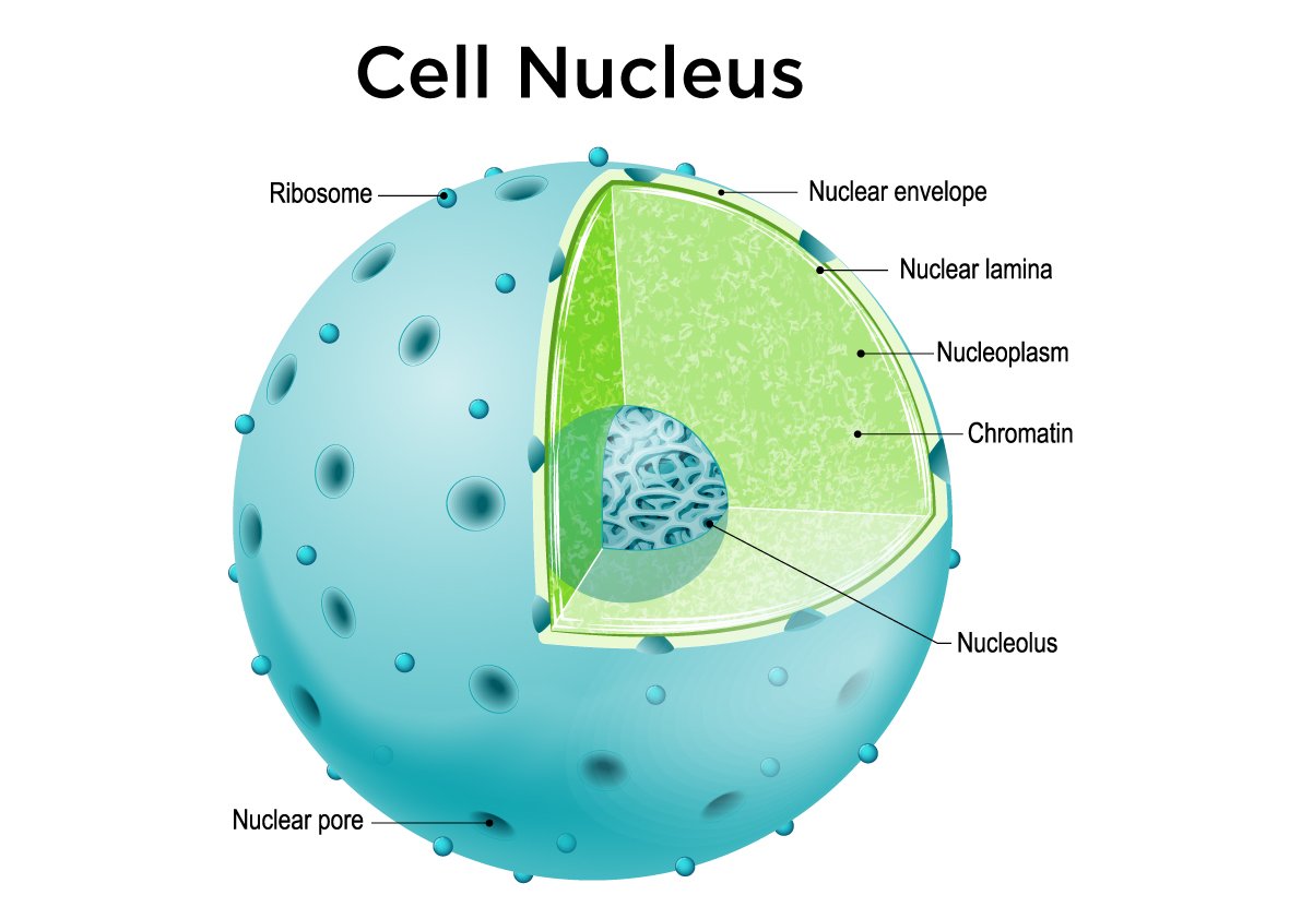
The nucleus is a membrane-bound organelle that contains the genetic material of the cell. It is often referred to as the control center of the cell because it regulates gene expression and controls cell growth and reproduction. The nucleus contains the DNA, which carries the instructions for making proteins and other molecules necessary for cell function.
Structure of Nucleus: The nucleus is surrounded by a double-layered nuclear membrane, also known as the nuclear envelope. It has a porous structure that allows for the movement of molecules between the nucleus and the cytoplasm. The nucleus also contains nucleoli, which are responsible for the production of ribosomes, and chromatin, which is the DNA and protein complex that makes up the chromosomes.
Functions of Nucleus:
- Stores and protects the genetic material of the cell
- Controls gene expression and regulates cell growth and reproduction
- Produces ribosomes, which are involved in protein synthesis
Cytoplasm
The cytoplasm is a jelly-like material that fills the space between the cell membrane and the nucleus. It contains various cellular organelles and structures, including the cytoskeleton, mitochondria, endoplasmic reticulum, Golgi apparatus, and ribosomes. The cytoplasm is responsible for supporting and protecting the organelles and providing a medium for cellular processes to occur.
Structure of Cytoplasm: The cytoplasm is a complex mixture of water, salts, and organic molecules. It is composed of a network of filaments called the cytoskeleton, which provides structural support for the cell and helps in cellular movement. The cytoplasm also contains various organelles suspended in the cytosol, which is the liquid portion of the cytoplasm.
Functions of Cytoplasm:
- Provides a medium for cellular processes to occur
- Supports and protects cellular organelles
- Facilitates cellular movement and shape changes
Cytoskeleton
The cytoskeleton is a network of protein filaments that provides structural support to the cell and helps in cellular movement. It is composed of three types of filaments: microfilaments, microtubules, and intermediate filaments.
Structure of Cytoskeleton: Microfilaments are the thinnest filaments, made up of actin protein. They are involved in cell contraction, cell division, and cell movement. Microtubules are larger hollow tubes made up of tubulin protein. They provide structural support and serve as tracks for intracellular transport. Intermediate filaments are intermediate in size and provide mechanical strength to cells and tissues.
Functions of Cytoskeleton:
- Provides structural support to the cell
- Helps in cellular movement and shape changes
- Facilitates intracellular transport
Lysosomes
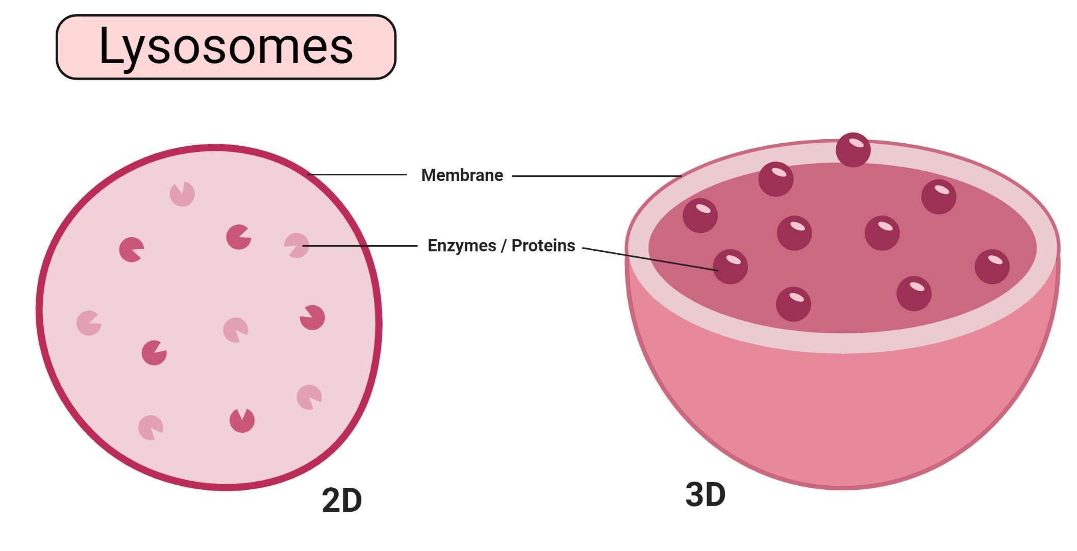
Lysosomes are membrane-bound organelles that contain digestive enzymes. They are involved in the breakdown of macromolecules, cell waste disposal, and cell renewal. Lysosomes play a crucial role in maintaining cellular homeostasis and removing damaged or unnecessary cellular components.
Structure of Lysosomes: Lysosomes are round organelles surrounded by a membrane. They contain various hydrolytic enzymes that break down macromolecules such as proteins, carbohydrates, and lipids into smaller molecules. The enzymes inside lysosomes are active in an acidic environment, which is maintained by proton pumps in the lysosomal membrane.
Functions of Lysosomes:
- Breaks down macromolecules into smaller molecules
- Removes damaged or unnecessary cellular components
- Plays a role in cell waste disposal and recycling
- Regulates cell renewal process
Nucleolus
The nucleolus is a distinct structure within the nucleus that is responsible for the production of ribosomes. It is involved in the synthesis and assembly of ribosomal RNA (rRNA) and ribosomal proteins. The nucleolus plays a crucial role in protein synthesis and cell growth.
Structure of Nucleolus: The nucleolus is not surrounded by a membrane but is composed of protein and RNA molecules. It is located within the nucleus and is often visible as a dark spot under a microscope.
Functions of Nucleolus:
- Produces ribosomes, which are involved in protein synthesis
- Assembles ribosomal RNA (rRNA) and ribosomal proteins
Mitochondria
Mitochondria are often referred to as the powerhouse of the cell because they are responsible for generating energy in the form of ATP (adenosine triphosphate). They are membrane-bound organelles that have their own DNA and can reproduce independently within the cell through a process called fission.
Structure of Mitochondria: Mitochondria have a double membrane structure. The outer membrane is smooth, while the inner membrane is highly folded, forming structures called cristae. The inner membrane encloses the mitochondrial matrix, which contains enzymes necessary for ATP production. Mitochondria also contain their own DNA, known as mitochondrial DNA (mtDNA).
Functions of Mitochondria:
- Generates energy in the form of ATP through cellular respiration
- Plays a role in cell signaling and apoptosis (programmed cell death)
- Regulates calcium ion levels in the cell
- Contains enzymes involved in lipid metabolism
Endoplasmic Reticulum (ER)
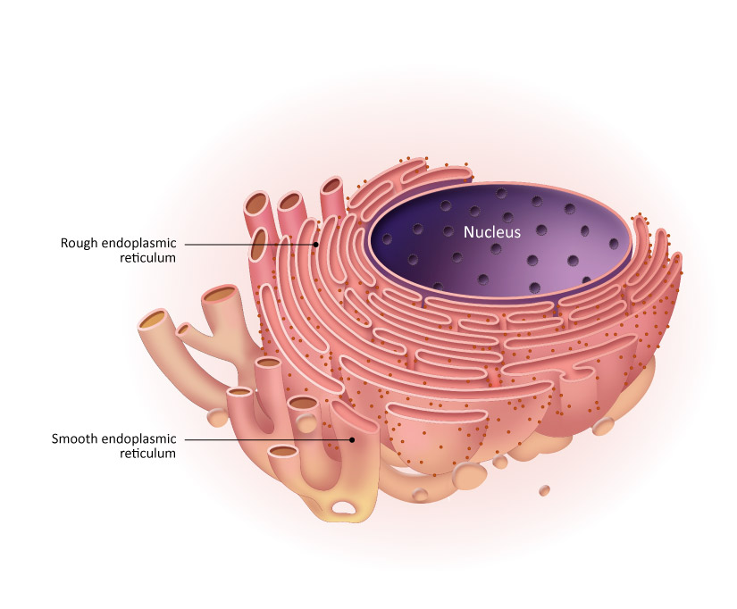
The endoplasmic reticulum (ER) is a complex network of membranous sacs, tubules, and cisternae. It is involved in the synthesis, folding, and transport of proteins and lipids. The ER is classified into two types: rough endoplasmic reticulum (RER), which is studded with ribosomes, and smooth endoplasmic reticulum (SER), which lacks ribosomes.
Structure of Endoplasmic Reticulum (ER): The ER is composed of a network of membranous sacs, tubules, and cisternae. The rough endoplasmic reticulum (RER) is studded with ribosomes, while the smooth endoplasmic reticulum (SER) lacks ribosomes. The RER is involved in protein synthesis and folding, while the SER is involved in lipid metabolism and detoxification.
Functions of Endoplasmic Reticulum (ER):
- Synthesizes proteins and lipids
- Modifies and packages proteins for transport within the cell
- Involved in calcium ion storage and release
- Detoxifies drugs and toxins
Ribosomes
Ribosomes are small organelles made up of RNA-rich cytoplasmic granules. They are involved in protein synthesis, where they read the messenger RNA (mRNA) and assemble amino acids into polypeptide chains.
Structure of Ribosomes: Ribosomes are composed of two subunits: a large subunit and a small subunit. Each subunit is made up of ribosomal RNA (rRNA) and proteins. Ribosomes can be found freely floating in the cytoplasm or attached to the endoplasmic reticulum.
Functions of Ribosomes:
- Synthesizes proteins by assembling amino acids into polypeptide chains
- Reads the genetic code from messenger RNA (mRNA)
Golgi Apparatus
The Golgi apparatus, also known as the Golgi complex or Golgi bodies, is involved in the processing, packaging, and sorting of proteins and lipids for transport within the cell or secretion outside the cell.
Structure of Golgi Apparatus: The Golgi apparatus is composed of flattened, stacked pouches called cisternae. It has three primary compartments: the cis Golgi network, the medial cisternae, and the trans Golgi network. The Golgi apparatus is closely associated with the endoplasmic reticulum and receives proteins and lipids from the ER for further processing.
Functions of Golgi Apparatus:
- Processes, modifies, and packages proteins and lipids
- Sorts proteins and lipids for transport within the cell or secretion outside the cell
- Plays a role in the synthesis of complex carbohydrates
Peroxisomes
Peroxisomes are small, membrane-bound organelles that contain enzymes involved in various metabolic reactions. They are involved in the breakdown of fatty acids, the detoxification of harmful substances, and the synthesis of certain lipids.
Structure of Peroxisomes: Peroxisomes are round organelles surrounded by a single membrane. They contain enzymes such as catalase and peroxidase, which are involved in metabolic reactions.
Functions of Peroxisomes:
- Breaks down fatty acids through beta-oxidation
- Detoxifies harmful substances, such as hydrogen peroxide
- Synthesizes certain lipids, such as plasmalogens
Centrosome
The centrosome is a small organelle located near the nucleus, which plays a crucial role in cell division. It is involved in the organization of microtubules and the formation of the mitotic spindle during cell division.
Structure of Centrosome: The centrosome consists of two centrioles, which are cylindrical structures composed of microtubules. The centrosome is surrounded by pericentriolar material, which contains proteins involved in microtubule organization.
Functions of Centrosome:
- Organizes microtubules and plays a role in cell division
- Forms the mitotic spindle, which helps in the separation of chromosomes during cell division
Vacuole
Vacuoles are membrane-bound organelles present in animal cells. They are involved in various functions, including maintaining cell shape, storing water and nutrients, and regulating osmotic pressure.
Structure of Vacuole: Vacuoles are membrane-bound sacs that contain water, ions, nutrients, and waste materials. In animal cells, vacuoles are smaller and less prominent compared to plant cells.
Functions of Vacuole:
- Maintains cell shape and rigidity
- Stores water, ions, nutrients, and waste materials
- Regulates osmotic pressure within the cell
Nucleopore
Nucleopores are tiny holes present in the nuclear membrane that allow the movement of nucleic acids and proteins across the nuclear membrane. They are involved in the transport of molecules between the nucleus and the cytoplasm.
Structure of Nucleopore: Nucleopores are complex structures composed of proteins and nucleic acids. They form a selective barrier that regulates the passage of molecules in and out of the nucleus.
Functions of Nucleopore:
- Regulates the movement of nucleic acids and proteins between the nucleus and the cytoplasm
- Facilitates the import and export of molecules involved in gene expression and protein synthesis
Cilia and Flagella
Cilia and flagella are hair-like structures that extend from the surface of animal cells. They are involved in cell movement and the movement of substances across the cell surface.
Structure of Cilia and Flagella: Cilia and flagella are composed of microtubules arranged in a 9+2 pattern. The 9+2 pattern refers to the arrangement of nine pairs of microtubules surrounding two central microtubules.
Functions of Cilia and Flagella:
- Facilitates cell movement
- Moves substances across the cell surface, such as mucus in the respiratory tract
Endosomes and Endocytosis
Endosomes are membrane-bound organelles involved in the sorting, recycling, and degradation of molecules that enter the cell through endocytosis. Endocytosis is a process by which cells take in substances from the external environment by engulfing them with the cell membrane.
Structure of Endosomes and Endocytosis: Endosomes are vesicles derived from the cell membrane through endocytosis. They contain various proteins and enzymes involved in the sorting and processing of molecules. Endocytosis involves the formation of vesicles that transport molecules from the cell membrane to the endosomes.
Functions of Endosomes and Endocytosis:
- Sorts, recycles, and degrades molecules taken in through endocytosis
- Plays a role in the regulation of cell signaling and membrane transport
Intermediate Filaments
Intermediate filaments are a type of cytoskeletal protein that provides mechanical support and strength to animal cells. They are involved in maintaining the structural integrity of cells and tissues.
Structure of Intermediate Filaments: Intermediate filaments are composed of various fibrous proteins, including keratins, vimentin, and neurofilaments. They form a network of filaments that helps to support and maintain the shape of the cell.
Functions of Intermediate Filaments:
- Provides mechanical support and strength to cells and tissues
- Maintains the structural integrity of the cell
Microfilaments
Microfilaments, also known as actin filaments, are the thinnest filaments of the cytoskeleton. They are involved in cell contraction, cell division, and cell movement.
Structure of Microfilaments: Microfilaments are made up of actin protein, which forms a network of filaments. They are often found near the cell membrane and play a crucial role in cell shape changes and movement.
Functions of Microfilaments:
- Facilitates cell contraction and movement
- Plays a role in cell division and shape changes
Microtubules
Microtubules are hollow tubes made up of tubulin protein. They are involved in various cellular processes, including cell division, intracellular transport, and cell shape maintenance.
Structure of Microtubules: Microtubules are composed of tubulin protein, which forms hollow tubes. They have a structure known as the 9+2 arrangement, where nine pairs of microtubules surround two central microtubules.
Functions of Microtubules:
- Provides structural support to the cell
- Facilitates cell division and chromosome separation
- Involved in intracellular transport of organelles and vesicles
Plasma Membrane
The plasma membrane, also known as the cell membrane, is a thin, semipermeable membrane that surrounds the animal cell. It plays a crucial role in maintaining cell homeostasis and regulating the exchange of molecules between the cell and its environment.
Structure of Plasma Membrane: The plasma membrane is composed of a lipid bilayer with embedded proteins. The lipid bilayer consists of two layers of phospholipids, with hydrophobic tails facing inward and hydrophilic heads facing outward. The proteins embedded in the lipid bilayer have various functions, including transport of molecules, cell signaling, and cell adhesion.
Functions of Plasma Membrane:
- Maintains cell homeostasis by regulating the exchange of molecules
- Controls the entry and exit of substances into and out of the cell
- Facilitates cell signaling and communication
- Provides structural support to the cell
Microvilli
Microvilli are small, finger-like projections on the surface of some animal cells. They increase the surface area of the cell, allowing for enhanced absorption and secretion.
Structure of Microvilli: Microvilli are composed of actin filaments, which are part of the cytoskeleton. They are found on the surface of cells that are involved in absorption, such as the cells lining the intestines.
Functions of Microvilli:
- Increase the surface area of the cell for enhanced absorption and secretion
- Facilitate the exchange of nutrients and waste products
Animal Cell Types
Animal cells can be classified into various types based on their structure and function. Some of the common types of animal cells include:
- Muscle Cells: These cells are involved in muscle contraction and movement.
- Nerve Cells: Also known as neurons, these cells transmit electrical signals in the nervous system.
- Epithelial Cells: These cells form the lining of body surfaces and cavities, providing protection and secretion.
- Connective Tissue Cells: These cells support and connect different tissues and organs in the body.
- Skin Cells: Also known as epithelial cells, these cells form the outer layer of the skin, providing protection and sensation.
- Blood Cells: These cells are involved in transportation of oxygen, nutrients, and waste products in the body.
- Fat Cells: Also known as adipocytes, these cells store energy in the form of fat and play a role in insulation and cushioning.
Animal Cell Size and Shape
Animal cells come in a variety of sizes and shapes, depending on their function and location in the body. The size of animal cells can range from a few micrometers to a few millimeters. The largest known animal cell is the ostrich egg, which can stretch over 5.1 inches across and weighs about 1.4 kilograms. On the other hand, the smallest animal cells are neurons, which are just 100 microns across.
Animal cells can have various shapes, including flat, oval, rod-shaped, curved, spherical, concave, and rectangular. The shape of the cell is determined by its function and the organization of its cytoskeleton. The absence of a cell wall in animal cells allows for greater flexibility and adaptability, allowing them to take on different shapes.
Animal Cells vs. Plant Cells: Difference Between Cells
Animal cells and plant cells share some similarities, as they are both eukaryotic cells with membrane-bound organelles. However, there are also significant differences between the two cell types.
One of the main differences is the presence of a cell wall in plant cells, which provides rigidity and support to the cell. Animal cells lack a cell wall, allowing for greater flexibility and adaptability. Instead of a cell wall, animal cells have a plasma membrane that controls the entry and exit of substances into and out of the cell.
Another difference is the presence of chloroplasts in plant cells, which are responsible for photosynthesis. Animal cells do not have chloroplasts, as they are not capable of photosynthesis and obtain their energy from other sources.
Plant cells also have larger vacuoles compared to animal cells. Vacuoles in plant cells play a role in maintaining cell shape, storing water and nutrients, and regulating osmotic pressure. Animal cells have smaller vacuoles that perform similar functions but to a lesser extent.
Animal cells and plant cells also differ in terms of their organelles. For example, animal cells have centrioles, which are involved in cell division, while plant cells do not. Plant cells also contain plastids, such as chloroplasts, which are responsible for photosynthesis.

How Does the Animal Cell Work?
Animal cells work together to carry out various functions necessary for life. Each organelle within the cell has a specific function and contributes to the overall functioning of the cell.
The cell membrane controls the entry and exit of substances into and out of the cell, maintaining cell homeostasis. The nucleus contains the genetic material of the cell and controls gene expression and cell growth. The cytoplasm provides a medium for cellular processes to occur and supports and protects cellular organelles.
The cytoskeleton provides structural support to the cell and helps in cellular movement and shape changes. Lysosomes break down macromolecules and remove damaged or unnecessary cellular components. The nucleolus produces ribosomes, which are involved in protein synthesis. Mitochondria generate energy in the form of ATP through cellular respiration.
The endoplasmic reticulum is involved in the synthesis, processing, and transport of proteins and lipids. Ribosomes are responsible for protein synthesis, while the Golgi apparatus processes, modifies, and packages proteins and lipids for transport or secretion. Peroxisomes are involved in various metabolic reactions, such as fatty acid breakdown and detoxification.
The centrosome plays a role in cell division, while vacuoles help maintain cell shape and store water and nutrients. Nucleopores regulate the movement of molecules between the nucleus and the cytoplasm. Cilia and flagella facilitate cell movement, and endosomes are involved in the sorting and processing of molecules.
The functioning of animal cells relies on the coordination and interaction of these organelles, allowing for the proper functioning of the cell and the organism as a whole.
Characteristics of Animal Cell
Animal cells share some common characteristics that distinguish them from other types of cells. Some of the key characteristics of animal cells are:
- Eukaryotic: Animal cells are eukaryotic cells, meaning they have a true nucleus and other membrane-bound organelles.
- Lack of Cell Wall: Animal cells do not have a cell wall, allowing for greater flexibility and adaptability compared to plant cells.
- Membrane-Bound Organelles: Animal cells have various membrane-bound organelles, such as the nucleus, mitochondria, and endoplasmic reticulum, which carry out specific functions within the cell.
- Heterotrophic: Animals cannot produce their own food and rely on the consumption of other organisms for energy and nutrients.
- Multicellular: Animals are multicellular organisms, meaning they are made up of more than one cell.
- Specialized Cell Types: Animal cells can differentiate into specialized cell types, such as muscle cells, nerve cells, and skin cells, to perform specific functions in the body.
How Kunduz Can Help You Learn Animal Cells?
Kunduz is your go-to resource for learning about animal cells and other scientific topics. We provide comprehensive and easy-to-understand educational materials, including articles, diagrams, and interactive learning tools. Whether you are a student, teacher, or simply curious about the world of biology, Kunduz is here to support your learning journey.